Shuai Liu1,2,
Jing Zhang3,
Xiao-Zhan Zhang1,
Hui-Hui Zhang1,
Xing-Wu Li1,
Shi-Jie Zhang1 ![]()
For correspondence:- Shi-Jie Zhang Email: zhangshijie2001@126.com Tel:+8637166295013
Received: 23 April 2016 Accepted: 14 August 2016 Published: 30 September 2016
Citation: Liu S, Zhang J, Zhang X, Zhang H, Li X, Zhang S. Triptolide induces cell apoptosis in human stomach cancer cell via caspase 3-dependent cascade pathway. Trop J Pharm Res 2016; 15(9):1853-1858 doi: 10.4314/tjpr.v15i9.6
© 2016 The authors.
This is an Open Access article that uses a funding model which does not charge readers or their institutions for access and distributed under the terms of the Creative Commons Attribution License (http://creativecommons.org/licenses/by/4.0) and the Budapest Open Access Initiative (http://www.budapestopenaccessinitiative.org/read), which permit unrestricted use, distribution, and reproduction in any medium, provided the original work is properly credited..
Purpose: To evaluate the effect of triptolide on the induction of cell apoptosis in human gastric cancer BGC-823 cells.
Methods: The cytotoxicity of triptolide was evaluated by 3-(4, 5-dimethylthiazol-2-yl)-2, 5-diphenyltetrazolium bromide (MTT) assay. The effect of triptolide on cell proliferation was measured using lactate dehydrogenase (LDH) assay. Cell apoptosis was determined by Annexin V/propidium iodide (PI) double-staining assay.
Results: MTT results indicate that triptolide significantly decreased cancer cell numbers in dose- and time-dependent manners in MTT assay. Data from LDH assay showed that triptolide markedly induced cytotoxicity in gastric cancer cells. Triptolide also remarkably induced both early and late apoptotic process in BGC-823 cells. In addition, the compound down-regulated the ex
Conclusion: Taken together, the findings strongly indicates that the pro-apoptotic activity of triptolide is regulated by caspase 3-dependent cascade pathway, and thus needs to be further developed for cancer therapy.
Introduction
Gastric cancer is now ranking the second leading cause of cancer-related deaths worldwide [1]. Surgery combined with chemotherapy is the current option for most patients diagnosed with advanced gastric cancer [2]. Due to the high rates of side effects and rapid therapeutic resistance, the efficacy of traditional chemotherapeutic drugs in the cancer treatment has declined [3]. Therefore, it is necessary and urgent to develop novel therapeutic agents which have higher efficacy but with limited side effects and low resistance.
Emerging evidences have shown that natural compounds from medicinal plants exhibit promising results in controlling various diseases, including cancers [4,5]. The root of Trypterigium wilfordii Hook L, also known as “Thunder of God Vine” is one traditional Chinese medicine that has been reported to have diverse biological activities including anti-arthritis, anti-Alzheimer and anti-cancer effects [6-8]. Triptolide is one of the active diterpene triepoxide compound identified from Trypterigium wilfordii Hook L. To date, triptolide has been found to exert potent immunosuppressive and anti-inflammatory properties [9]. Triptolide is also demonstrated to have anti-cancer effects in several cancer types including lung cancer [10], liver cancer [11], pancreatic cancer [12], and colon cancer [13]. In addition, triptolide is also able to cause pancreatic cancer cell death through induction of apoptosis via inhibition of HSP70 [12]. Triptolide has also been demonstrated to abrogate cell growth of colon cancer and induce cell cycle arrest through inhibiting transcriptional activation of E2F [14]. A recent study also suggests that the induction of cell death by triptolide is modified by autophagy in cardiomyocytes [15].
In order to extend the usage of triptolide for cancer treatment, we will evaluate the potent cytotoxic effects of triptolide in human gastric BGC-823 cells, and also explore the possible underlying mechanisms of this action in the current study.
Methods
Chemicals and reagents
Triptolide, 3-(4,5-dimethylthiazol-2-yl)-2,5-diphenyltetrazolium bromide (MTT), dimethyl sulfoxide (DMSO), and propidium iodide (PI) were purchased from Sigma Chemical Co. (St. Louis, MO, USA). All other reagents, supplements for cell culture and assay kits were obtained from Life Technologies (NY, USA), otherwise stated.
Cell culture
BGC-823 human gastric cancer cells were purchased from the Cell Storehouse of the Chinese Academy of Science (Shanghai, China). The cells were maintained in DMEM medium supplemented with 10 % FBS and 1 % PS, and incubated in a humidified incubator at 37 °C with 5 % CO2.
Cell viability assay
In order to investigate the anti-cancer actions of triptolide, MTT assay was performed in BGC823 gastric cancer cells. The cells at a density of 5 × 10⁴ cells/mL were seeded in 96-well plates and treated with various concentrations of triptolide (10, 20, and 40 nM) or vehicle solution (0.1 % DMSO) for 48 or 72 h. Then, 20 μL of MTT (5 mg/mL) solution was added into each well and incubated for another 4 h. After replacing the culture medium with 100 μL of DMSO, the optical density (OD) was collected at the wavelength of 540 nm using a microplate reader (TECAN, Austria).
LDH leakage assay
Cell cytotoxicity was tested by LDH leakage assay according to the manufacturer’s instructions. Briefly, after triptolide treatments, cell-free supernatants (50 μL) were collected from each well and transferred into a new 96-well plate, and gently mixed with 50 μL of CytoTox 96® non-radioactive cytotoxicity assay reagents. The plate was kept in the dark at room temperature for 30 min. Stop solution (50 μL of each well) was added into the wells, before the OD value was examined at 492 nm using a microplate reader (TECAN, Austria).
Annexin V/PI double-staining assay
Annexin V/PI double-staining assay and flow cytometry (FACSalibur, BD) were performed to quantify the apoptotic cells after triptolide treatment. In brief, BGC-823 cells were seeded in 6-well plates at a density of 1 × 10⁵ cells/mL and treated with different concentrations of triptolide or DMSO for 48 or 72 h. The cells were then harvested, rinsed twice with PBS, resuspended in binding buffer and incubated with fluorescein isothiocyanate (FITC)-conjugated Annexin V (5 μL) and PI (5 μL) in the dark for 15 min at room temperature. Then, the cells were washed with PBS twice, before subjecting to flow cytometry for data collection.
Quantitative real-time polymerase chain reaction (q-PCR)
Total RNA was extracted with TRIzol® reagent, according to the manufacturer’s instructions. One microgram of RNA from each sample was used for the reverse transcription reaction using Oligo dT (18T) (Omega, NY, USA). The cDNA products were amplified for bcl-2, bax, and gapdh gene expression via qRT-PCR using specific primers. PCR was performed with SYBR Green PCR Master Mix using a 7900HT qPCR system thermal cycler (Applied Biosystems, CA). The Ct values for each sample were normalized to GAPDH mRNA, which was used as an internal control. Data from three independent experiments were collected and calculated.
The primers used for q-PCR were: bax, forward (5′-TGGAGCTGCAGAGGATGATTG-3′) and reverse (5′-GAAGTTGCCGTCAGAAAACATG-3′); bcl-2, forward (5′-CTGCACCTGACGCCCTT CACC-3′) and reverse (5′-CACATGACCCCACC GAACTCAAAGA-3′); GAPDH, forward (5′-AACGGGAAGCTTGTCATCAATGGAAA-3′) and reverse (5′-GCATCAGCAGAGGGGGCAGAG-3′)
Assessment of caspase-3 activity
After treatment with triptolide, caspase-3 activity was assessed using a commercial colorimetric activity assay kit (Beyotime, China), according to the manufacturer’s instruction.
Statistical analysis
Data are expressed as mean ± standard deviation (SD, n = 3). One-way ANOVA analysis was performed for statistical analysis using GraphPad Prism software, version 6.0. In all comparisons, p < 0.05 was considered statistically significant.
Results
Triptolide decreased cell proliferation in human gastric cancer BGC-823 cells
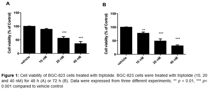
As shown in , treatment with triptolide (10, 20 and 40 nM) significantly decreased cell proliferation and cell viability in both time- and concentration-dependent manner. Cell viability of BGC-823 cells were inhibited by 44 and 64 % with triptolide treatment of 20 and 40 nM, respectively, for 48 h, and 51 and 69 % respectively for 72 h.
Triptolide induced cell cytotoxicity in human gastric cancer BGC-823 cells
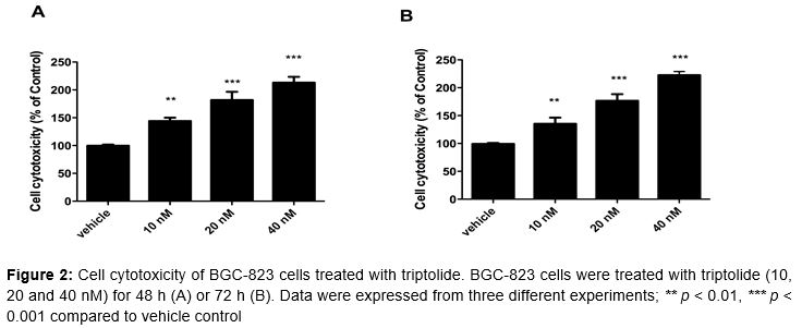
The cytotoxicity was examined by LDH leakage assay. Our results showed that toxic effects were increased in BGC-823 cells by triptolide treatment (10, 20, and 40 nM) in a dose-dependent manner ().
Triptolide induced cell apoptosis in human gastric cancer BGC-823 cells
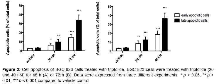
Cell apoptosis was measured by Annexin V and PI double-staining assays using flow cytometry. As shown in , the total apoptotic cells were increased 10.3 and 42.6 % by challenging with 20 nM and 40 nM of triptolide for 48 h (A), 14.5 and 48.3 % for 72 h (B), respectively. These results suggested triptolide significantly increased both early apoptosis and late apoptosis in a dose-and time-dependent manner.
Triptolide modulated the expression of apoptosis-related genes
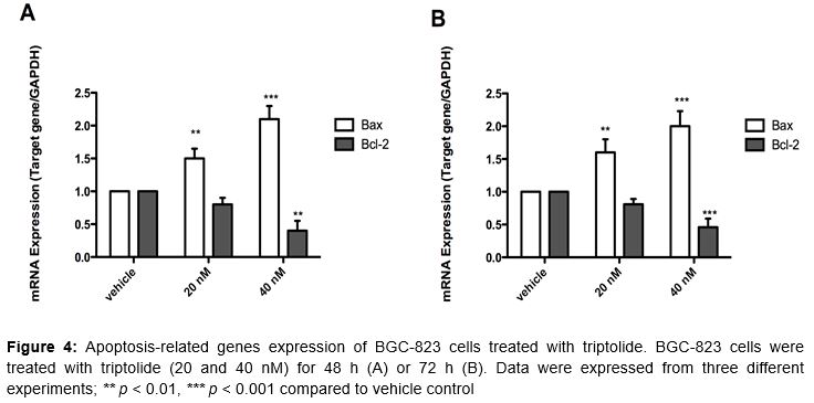
In order to study the underlying mechanisms for triptolide-induced apoptosis, apoptosis related genes including Bax and Bcl-2 mRNA were quantified by qPCR. As shown in , the expression of Bax mRNA was markedly up-regulated by triptolide treatment (20 and 40 nM) for 48 and 72 h, with over-expression of 50-110 %. However, the expression of Bcl-2 mRNA was significantly inhibited by triptolide (40 nM) for both 48 h and 72 h, with inhibitory rate of over 50 %.
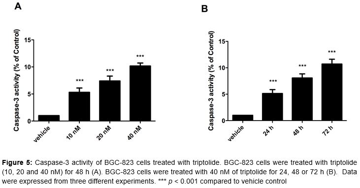
Triptolide activated caspase-3 activity in human gastric cancer BGC-823 cells
To confirm the involvement of caspase-3 in triptolide-induced cell apoptosis, caspase-3 activity was measured by colorimetric activity assay. The results showed that caspase-3 activity was increased by 5.3, 7.4 and 10.2 times by treatment with 10, 20 and 40 nM of triptolide in BGC-823 cells for 72 h (A). The activation of caspase-3 activity was also increased by triptolide (40 nM) in a time-dependent manner (B).
Discussion
Triptolide is one of the active ingredients of Tripterygium wilfordii, which possesses a broad spectrum of biological activities such as anti-inflammatory, anti-neoplastic and anti-cancer effects [15]. Triptolide exerts potent anti-cancer effects in several cancer types including lung cancer [10], liver cancer [11], pancreatic cancer [12], and colon cancer [13]. In our present study, we demonstrated that triptolide exhibited promising anti-cancer potential in human gastric BGC-823 cells. In addition, we also elucidated that the anti-cancer action of triptolide might be due to the induction of cell apoptosis in gastric cancer and the involvement of activation of caspase-3 pathway.
In our study, we used different method to evaluate the effects of triptolide in gastric BGC-823 cells including MTT, LDH leakage, Annexin V and PI double staining assays. The MTT assay can detect both normal cells and necrotic cells. Therefore, we further used LDH leakage assay to detect the cytotoxic effects of triptolide in BGC cells. In order to confirm the pro-apoptotic effects induced by triptolide, we performed flow cytometry with Annexin V and PI double staining. Our results showed that triptolide significantly increased LDH in the cultured medium and increased apoptotic cells in flow cytometric analysis, which strongly suggested that triptolide was able to induce both cytotoxic and pro-apoptotic effects in human gastric BGC-823 cells.
Cancer is fundamentally a disease of dysregulation of tissue growth, in which programmed cell death is interfered with and blocked by tissue microenvironment. Apoptosis is the main form of programmed cell death that maintains tissue and cell hemostasis [16]. During the development of apoptosis, both anti-apoptotic proteins such as Bcl2 and pro-apoptotic proteins such as Bax are involved in the initiation and modulation of the process of apoptosis [17]. The expression of Bcl2, Bax or the ratio of Bcl2 and Bax are normally used as index of apoptosis [18]. In our current study, we observed that triptolide induced a remarkable up-regulation of Bax mRNA and a significant decrease of Bcl2 mRNA, which consequently lead to an increase of the Bax/Bcl2 ratio.
These results indicate that triptolide-induced apoptosis employs both anti-apoptotic and pro-apoptotic proteins. The mitochondrial apoptotic pathway includes two alternate pathways, typically caspase-8 and caspase-9. Caspase-3 is a downstream molecule in caspase-9 pathway [19]. In our study, treatment with triptolide significantly stimulated the activation of caspase-3, suggesting that a caspase-9 pathway may be involved in the triptolide-induced cell apoptosis in gastric cancer.
Conclusion
Treatment with triptolide remarkably induces both early and late apoptotic process. Triptolide also down-regulates the expression of anti-apoptotic B-cell lymphoma-2 (bcl-2) and up regulates the expression of pro-apoptotic BCL-2-associated X (bax) in a dose-dependent manner. This process is accompanied by activation of caspase-3 in BGC-823 cells using caspase-3 activity assay. Thus, these findings indicate that the apoptotic activity of triptolide is probably regulated by caspase-3-dependent cascade way. The demonstrated pro-apoptotic effects of triptolide will shed some light on application of this drug in cancer treatment especially in gastric cancer.
Declarations
Acknowledgement
References
Archives


News Updates